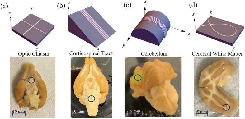
Fig. 2.
(Top row) Renderings of fiber geometries investigated in this study, and (bottom row) photographs of corresponding ferret brain ROIs. (a) Crossing fibers and optic chiasm, (b) inclined fibers and CST, (c) curving fibers out of plane and cerebellum, and (d) curving fibers in plane and cerebral white matter region.

