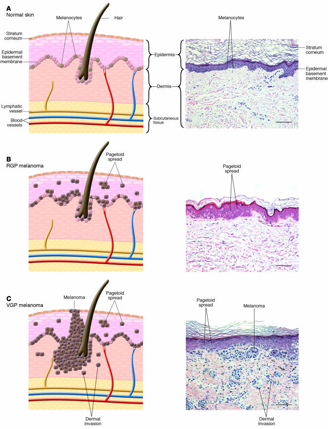
Figure 1.
Phases of histologic progression of melanocyte transformation. H&E-stained histologic sections and corresponding pictorial representation. (A) Normal skin. There is even distribution of normal dendritic melanocytes in the basal epithelial layer. (B) RGP in situ melanoma. Melanoma cells have migrated into the upper epidermis (pagetoid spread) and are scattered among epithelial cells in a “buckshot” manner. Cells have not penetrated the epidermal basement membrane. Melanoma cells show cytologic atypia, with large abundant cytoplasm and increased overall size compared with normal melanocytes. Nuclei are enlarged and hyperchromatic. Commonly, there is more junctional melanocytic hyperplasia (nests of tumor cells at the basement membrane zone) in RGP melanoma than portrayed in the histologic example. (C) VGP malignant melanoma. Melanoma cells show pagetoid spread and have penetrated the dermal-epidermal junction. Melanoma cells show cytologic atypia. Cells in the dermis cluster or individually invade. Magnification, ×20. Scale bar: 20 μm.

