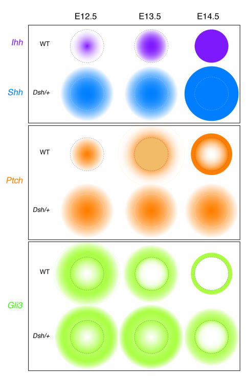
Figure 3.
Disrupted expression boundaries in Dsh/+ animals lead to perichondrial and joint defects (1). A transverse schematic of the phalangeal skeletal condensation, showing expression domains corresponding to a specific Hedgehog gene (Ihh in WT, Shh in Dsh/+), the Hedgehog target Ptch, and the Hedgehog repressor Gli3. Dotted lines indicate the normal cartilage-perichondrium boundary. In WT phalanges, Ihh (purple) is initially expressed (E12.5) in a small domain that expands over time to encompass the skeletal condensation. At E12.5 in Dsh/+ phalanges, Shh expression (blue) encompasses the normal Ihh domain and over time extends beyond what would normally be the cartilage-perichondrium boundary. Ptch (orange), an indicator of Hedgehog responsiveness, is first expressed in WT phalanges coincident with Ihh; eventually, Ptch is downregulated in cells expressing Ihh and upregulated in cells adjacent to the Ihh domain, where it functions to limit the spread of Hedgehog expression. Note the absence of color within the element, which shows the relative lack of Ptch in this domain (10). In Dsh/+ phalanges, Ptch is expressed coincident with the broader domain of Shh; however, in contrast to what occurs in WT tissues, Ptch does not become downregulated in chondrocytes. Gli3 (green), a repressor of Hedgehog signaling, is initially expressed in WT phalanges in a domain overlapping with Ihh; over time, this domain is restricted to the perichondrium, where Gli3 limits the activity of the Hedgehog protein. In Dsh/+ phalanges the Gli3 expression domain is initially broader, and only after an extended period of time does it become restricted to the perichondrium.

