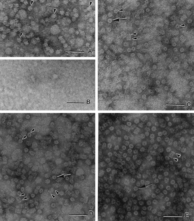
FIG. 3.
Transmission electron micrographs of negatively stained M. thermophila 20S proteasomes and subunits. (A) α subunit produced in E. coli independently of the β prosubunit. Seven-membered ring structures (α7) with no apparent central pore are visible (arrowheads). (B) β prosubunit produced in E. coli independently of the α subunit. (C) Authentic 20S proteasome purified from M. thermophila. End-on views (single arrow and arrowheads) and lateral views (double arrowheads) of the cylindrical 20S proteasome comprised of four stacked (α7β7β7α7) rings. (D) 20S proteasome produced in E. coli. The structures are indistinguishable from those in C. (E) The 20S proteasome assembled in vitro from α7 and β prosubunits independently produced in E. coli. The structures are indistinguishable from those in C and D. Bars, 50 nm.

