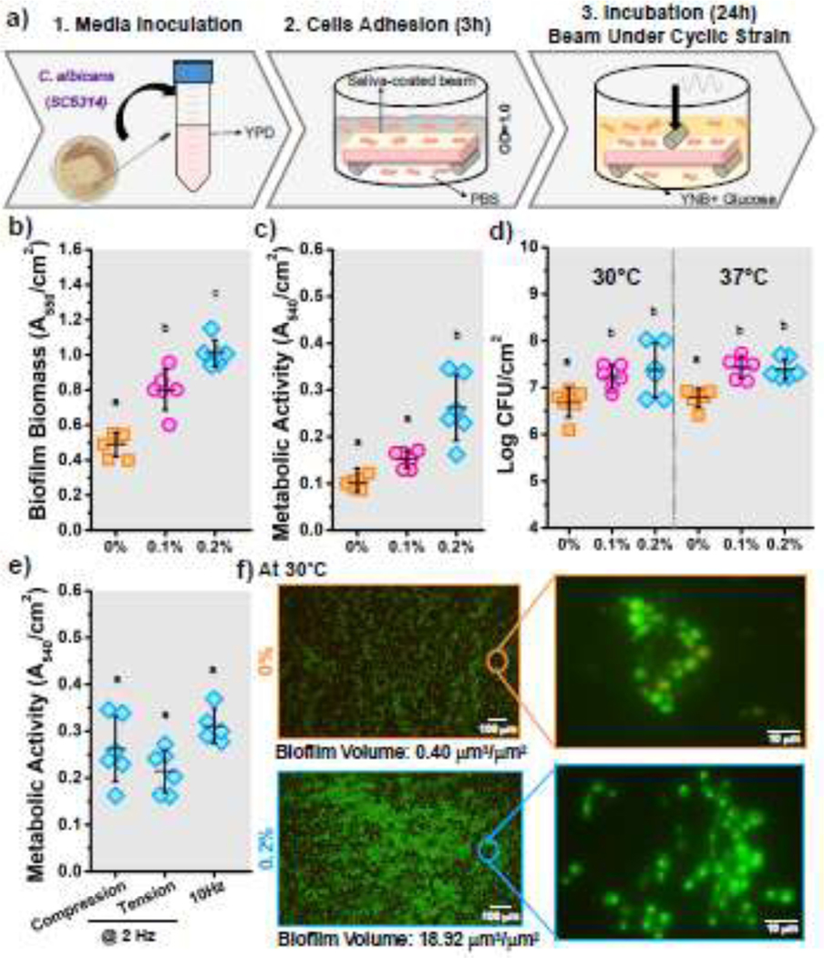
Figure 2.

C. albicans biofilm–biomaterial interactions. (a) Schematic of the biofilm model used to cultivate C. albicans biofilms on PMMA samples under cyclic mechanical strain. b) Biofilm biomass, c) metabolic activity, and d) number of viable cells of biofilms cultivated under different cyclic strain conditions (0%, 0.1%, and 0.2%). e) Metabolic activity of C. albicans biofilms grown on surfaces subjected to different types of strain (tension or compression) and with different frequencies (2 Hz and 10 Hz). f) Representative fluorescence microscopy images of C. albicans biofilms on PMMA surfaces under different repetitive strains. Samples were stained with SYTO 9 (green) and propidium iodide (red) to indicate live and dead fungi, respectively. N=6 samples for each evaluation. Means with different letters are significantly different from each other (p ≤ 0.05).
