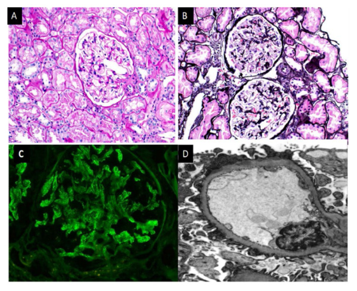
Figure 2.
Microscopic features of NELL-1 associated MN. A-Glomerulus on Periodic Acid Schiff stain, showing non-proliferative pattern and prominent capillary loops. B-Spikes and holes along the glomerular capillary loops seen on silver stain. C-Segmental and incomplete global IgG staining in glomeruli on immunofluorescence microscopy. D-Electron-dense deposits along the glomerular capillary loops on Electron microscopy.

