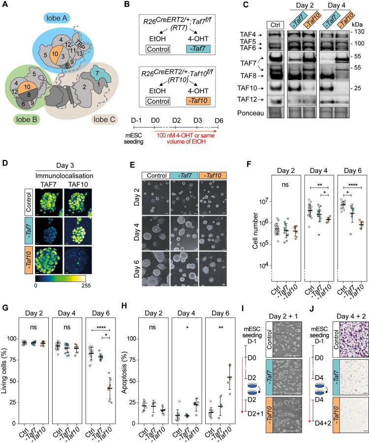
Figure 1: Phenotypic analysis of the conditional depletion of TAF7 or TAF10 in mESCs.
(A) Schematic structure of TFIID. Lobe A is indicated in blue, lobe B in green and lobe C in beige. (B) Strategy of the induction of the deletion of Taf7 (-Taf7) in R26CreERT2/+;Taf7f/f (RT7) and Taf10 (-Taf10) in R26CreERT2/+;Taf10f/f (RT10) mESCs. Cells are plated at day (D) −1, 100 nM 4-hydroxytamoxifen (4-OHT, depleted) or the same of volume of ethanol (control) were added at D0 and maintained until the day of the analysis. (C) Western blot analyses of TAF4, TAF5, TAF6, TAF7, TAF8, TAF10 and TAF12 proteins expression after Taf7 (-Taf7) or Taf10 (-Taf10) deletions after 2 and 4 days of 4-OHT treatment. As a control (Ctrl), RT7 cells were treated 2 days with EtOH. The Ponceau staining is displayed at the bottom of the panel. (D) Immunolocalization of TAF7 and TAF10 in RT7 and RT10 cells treated for 3 days with 4-OHT. As a control, RT10 cells were treated with EtOH for 3 days. Color scale (Green Fire Blue LUT scale) is indicated at the bottom. Scale bar; 15μm. (E) Monitoring of cell growth over time. RT7 and RT10 cells were treated over 6 days, and pictures were taken at D2, D4 and D6. Scale bar; 50 μm. (F) Log10 of the total number of cells at D2, D4 and D6 of treatment. (G) Percentage of living cells after 2, 4, and 6 days of treatment determined by Trypan blue staining. For (E, F), Ctrl: D2; N = 20, D4; n = 20, D6; n = 15, -Taf7: D2; n = 13, D4; n = 13, D6; n = 8, -Taf10: D2; n = 7, D4; n = 7, D6; n = 7 biological replicates. (H) Percentage of apoptotic cells after 2, 4 and 6 days of treatment determined by Annexin V and propidium iodide (PI) staining. For D2, D4 and D6, Ctrl; n = 9, -Taf7; n = 4, -Taf10; n = 5 biological replicates. The bars correspond to the mean ± standard deviation. Kruskal-Wallis test followed by Dunn post hoc test if significant: ns; not significant, * <0.05; **<0.01; *** <0.001, **** <0.0001. (I) Cell density after passage at D2 and 1 day of extra culture. (J) Cell density evaluated by Crystal violet staining after passage at D4 and 2 days of culture. Scale bar; 150 μm. For D, F and G, the control conditions correspond to RT7 cells treated with EtOH.

