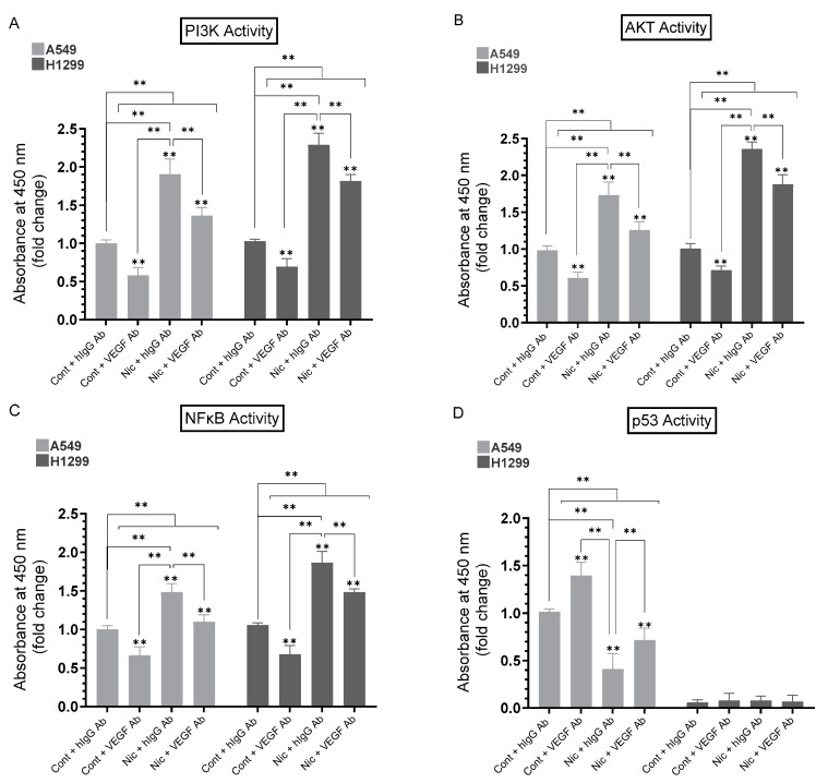
Figure 6.
Anti-VEGF antibodies decreased the activities of PI3K, AKT, and NFκB in A549 and H1299 cells without or with nicotine and increased p53 activation in A549 cells. Cells were grown in media with 10% FBS for 24 h, serum-starved overnight, then incubated in serum-free media for 72 h without or with nicotine (Nic, 1 µM), hIgG (20 μg/mL) as a control, anti-VEGF-specific antibodies (20 μg/mL), or in combination. The activities of PI3K, AKT, NFκB, and p53 were measured (Section 2). Data were averaged and expressed as a fold change relative to the control cells treated with hIgG (Cont + hIgG Ab) of each cell line (A–C) or to A549 (Cont + hIgG Ab) (D) using the GraphPad 9.5.1 software (n = 5). Asterisks indicate a statistically significant difference from the control of each cell line. Statistical differences between different groups were analyzed by an ordinary one-way analysis of variance (ANOVA) followed by Tukey’s post hoc multiple comparison test, ** p < 0.01.

