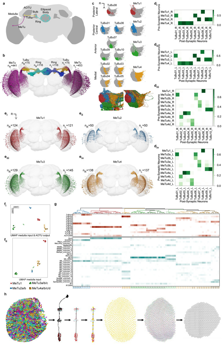
Fig. 1 |. Identification and classification of MeTu neurons within the Anterior Visual Pathway.

a, Diagram of the Drosophila melanogaster central brain, emphasizing the Anterior Visual Pathway (AVP). Important regions are darker gray, including medulla, AOTU, mulb, and EB (the former three have counterparts in both hemispheres). The three crucial neurons of the AVP are MeTu (from the Medulla to the AOTU_SU, purple), TuBu (from the AOTU_SU to the bulb, yellow), and Ring/ER (from the bulb to the EB, cyan). b, All MeTu (n=451 on the left, n=441 on the right), TuBu (n=75 on the left, n=75 on the right), and visual Ring (n=116 on the left, n=116 on the right) neurons. c, Top: synapse plots of TuBu (left) and MeTu (right) neurons in the posterior lateral (red), posterior central (blue), anterior (green), and medial (yellow) region of the AOTU_SU. Bottom: all TuBu (left) and MeTu (right) in the AOTU_SU_R. di-ii, Synaptic weight matrix of MeTu type to TuBu type connectivity in the right (di) and left (dii) hemisphere. diii-iv, Synaptic weight matrix of MeTu subtype to TuBu type connectivity in the right (diii) and left (div) hemisphere. ei-iv, All neurons of types MeTu1 (ei), MeTu2 (eii), MeTu3 (eiii), and MeTu4 (eiv). fi-ii, UMAPs of all MeTu neurons with identified upstream partners based on the synaptic weight of the top 5 medulla input neuron types and AOTU output neuron types (fi) or just synaptic weight of the top 5 medulla input neuron types (see Methods for details)(fii). Groupings are generally consistent with MeTu1-4 groups in the main text with a notable exception that MeTu3a neurons are closer to MeTu2 neurons (because of the similar polarization input) than other MeTu3 neurons. g, Synaptic weight matrix of all MeTu neurons with identified upstream partners (columns) and their AOTU output partners (top rows in red) and top5 medulla input neuron types (bottom rows in dark cyan). Dendrogram branches and column labels are color-coded according to MeTu. h, Process of defining medulla columns and layers from all Mi1 neurons, a uni-columnar cell type, shown for the right optic lobe. From left to right: Render of all Mi1neurons of the right optic lobe, a single Mi1 neuron with pre- (red) and postsynaptic (cyan) sides, distal-proximal axis of a column is given by PC1 of a PCA on all synaptic sides of the corresponding Mi1 neuron, defining layer markers based on the upper and lower bound of the distal-proximal axis, m6 layer marker of all columns, manual assignment of neighboring columns along the horizontal (black), vertical (red), p (blue) and q (green) axis, and the resulting hex grid.
