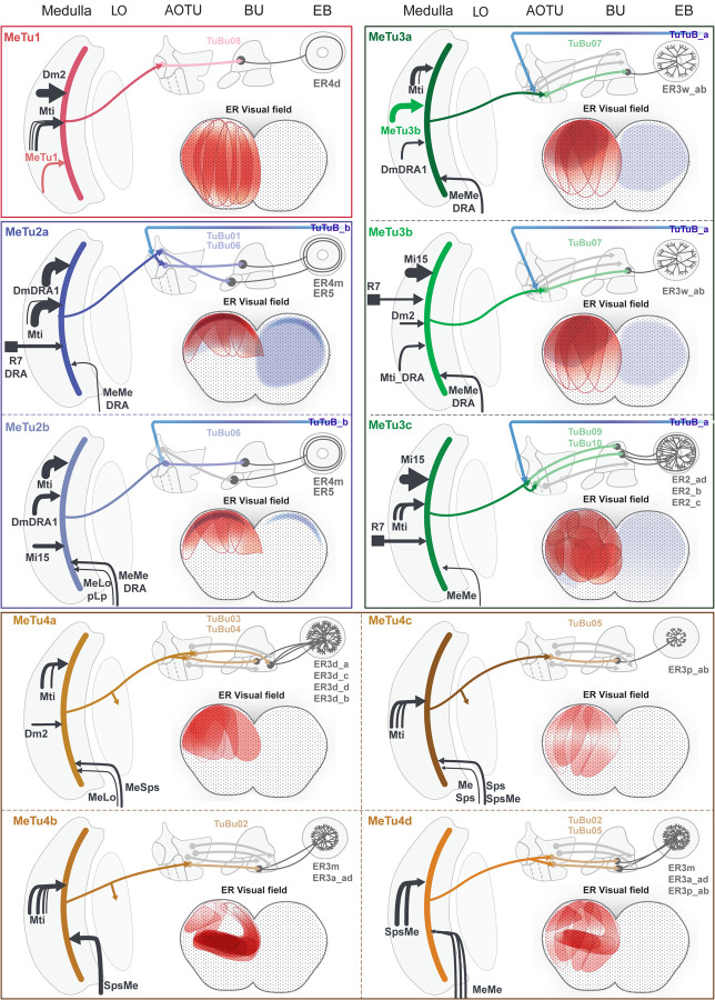
Fig. 7 |. Overviews of Parallel Anterior Visual Pathways.
Each panel shows an generalized neural pathway and receptive field of a MeTu subtype. From the left to top right side, there is a diagram of the pathway from the medulla to the EB. Medulla inputs to MeTu are shown on the left of the medulla if they come from the retina or medulla, and on the right if they come from the central brain. Photoreceptor inputs in the retina are shown as squares. The AVP from the MeTu to the TuBu and Ring neurons are shown, as well as if the MeTu has outputs in the lobula or synapses with TuTuB neurons in the AOTU. Receptive fields of the relevant Ring neurons are on the bottom right. Red indicates excitatory input from MeTu neurons, whereas blue indicates putative inhibitory input from TuTuB neurons. Only inhibitory input from TuTuB is shown; AOTU046 is not included because the excitatory/inhibitory nature of the neuron is unknown [65]. The interaction between AVP channels appears to be minimal. In other words, direct interaction between the four major MeTu types is negligible downstream of the medulla.

