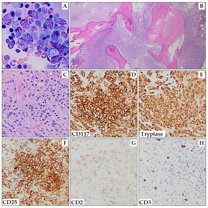
Figure 2.
Aggressive systemic mastocytosis. Aspirate smears show areas with numerous hypogranular and/or spindle-shaped MCs (A; 200×). The marrow shows multifocal interstitial and paratrabecular aggregates of MCs, as well as a few small areas of residual trilineage hematopoiesis (B; 20×). One highlighted MC aggregate (C) shows expression of CD117 (D), tryptase (E), CD25 (F), and CD2 (weak, partial; (G)). CD3 (H) is negative in MCs. (C–H) are all at 100× magnification.

