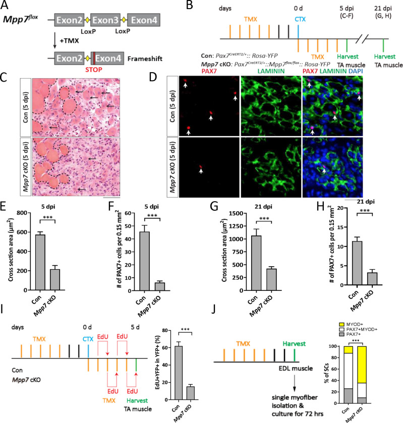
Figure 1. Mpp7 cKO in Pax7+ MuSCs shows defects in regeneration and MuSC self-renewal.
(A) Diagram of Mpp7 floxed allele (Mpp7flox; loxP, yellow diamond) for tamoxifen (TMX) inducible Cre-mediated cKO. After recombination, out of-frame (Frameshift) joining of Exons 2 and 4 introduces an early stop codon (STOP).
(B) Regimen of TMX administration, cardiotoxin (CTX) injury, and tibialis anterior (TA) muscle harvest; d, day; dpi, days post injury. Genotypes of control (Con) and Mpp7 cKO are indicated.
(C-F) Mpp7 cKO regeneration defects at 5 dpi. Representative images of H&E histology are in (C), immunofluorescence (IF) for PAX7 and LAMININ in (D, with DAPI), and quantification of regenerated myofiber cross sectional area in (E) and of PAX7+ MuSC density in (F). Black arrows indicate regenerated myofibers; dashed lines, boundary of injury; white arrows, PAX7+ MuSCs. N = 5 mice, each.
(G, H) Quantifications of regenerated myofiber cross sectional area (G) and PAX7+ MuSC density (H) at 21 dpi. N = 5 mice, each.
(I) Regimen of in vivo EdU incorporation to assess the percentage of proliferated YFP-marked cells at 5 dpi; quantification to the right. N = 5 mice, each.
(J) Regimen of cell fate determination using single myofiber culture. Cell fates were assessed by IF of PAX7 and MYOD; quantification to the right; keys to cell fates at the top. N = 3 Con mice, of total 601 cells; N = 4 Mpp7 cKO mice, of total 517 cells.
Data information: Scale bars = 25 μm in (C-D). (E-I) Error bars represent means ± SD; Student’s t-test (two-sided). (J) Chi-square test. ***, P<0.001.

