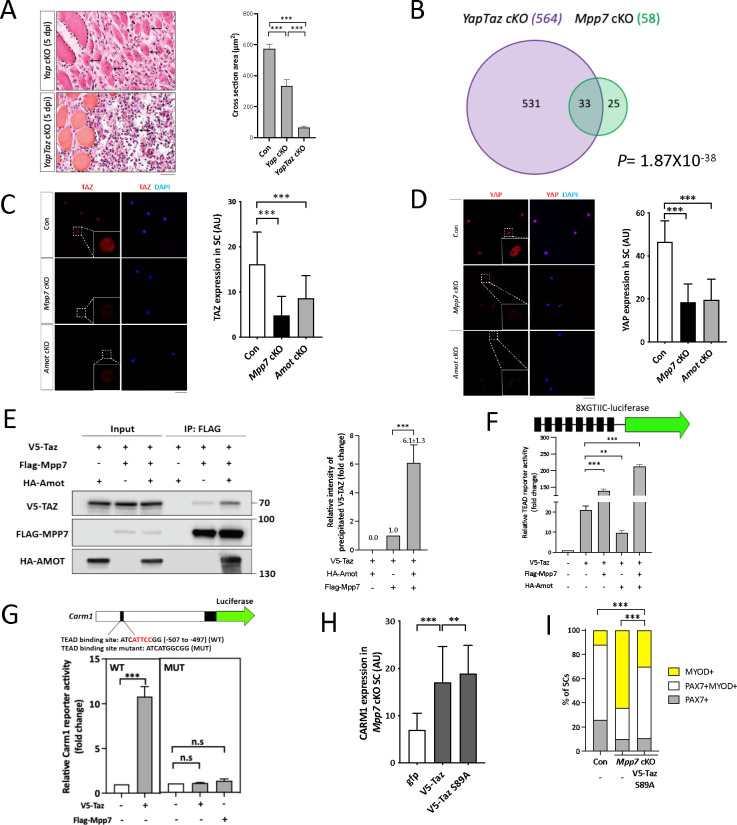
Figure 4. Mpp7/Amot regulatory network intersects with that of Yap/Taz.
(A) Representative H&E histology of Yap cKO and YapTaz cKO muscles at 5 dpi (Con histology not included); quantifications of regenerated myofiber cross sectional area to the right. N = 5, each genotype.
(B) Venn diagram shows overlapping DEGs between the YapTaz cKO and the Mpp7 cKO.
(C, D) Representative IF images of TAZ (C) and YAP (D) in FACS-isolated and cultured Con, Mpp7 cKO, and Amot cKO MuSCs at 48 h; quantified fluorescent signals (AU) to the right of corresponding images; 200 MuSCs from 2–3 mice in each group.
(E) Co-IP of V5-TAZ and HA-AMOT by FLAG-MPP7 expressed in 293T cells. Expression constructs and tagged epitopes for detection are indicated; (−), empty vector. Quantification of relative levels of co-IPed V5-TAZ is to the right. N = 3.
(F) Relative TEAD-reporter (8XGTIIC-luciferase, depicted at top) activities when co-transfected with V5-Taz, Flag-Mpp7, and/or Ha-Amot expression constructs in 293T cells; (−), empty vector. N =3.
(G) Relative activities of WT and TEAD-binding site mutated (MUT) Carm1-reporters co-transfected with V5-Taz or Flag-Mpp7 expression constructs; (−), empty vector. N = 3.
(H) Quantified IF signals (AU) of CARM1 in Mpp7 cKO MuSCs transfected with gfp (as control), V5-Taz WT and V5-Taz S89A expression constructs; 200 MuSCs from 2–3 mice in each group.
(I) Comparison of cell fate fractions among Con, Mpp7 cKO, and Mpp7 cKO MuSCs transfected with V5-Taz S89A expression construct in single myofiber culture; (−), empty vector; keys at the top; ≥ 217 cells in each group.
Data information: Scale bar = 25 μm in (A) and 50 μm in (C, D). Error bars represent means ± SD. Hypergeometric test was used in (B); One-way ANOVA with Tukey’s post hoc test was performed in (A, C, D, F-H); Chi-square test in (I). n.s, P > 0.05; **, P < 0.01; ***, P < 0.001.

