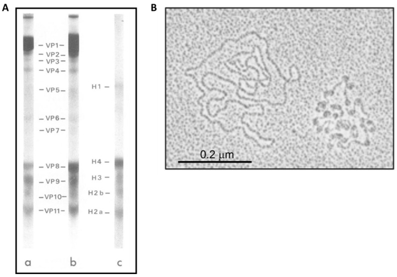
Figure 4.
HPV genome organization. (A) SDS-polyacrilamide cylindrical gel electrophoresis and gradient slab gel electrophoresis of SDS-dissociated papillomaviruses. Samples included highly purified BPV (a), HPV (b) proteins, and calf liver histones (c); (B) electron microscopy of HPV genomes showed naked HPV DNA molecules (left) and nucleoprotein-DNA complexes (right) [116]. (viral protein 1–11, VP 1–11; H1, histone 1; H2A, histone 2A; H2B, histone 2B; H3, histone 3; H4, and histone 4) [125].

