
Figure 4. TRPV4 regulates matrix stiffness dependent activation of VEGFR2 through YAP activation.
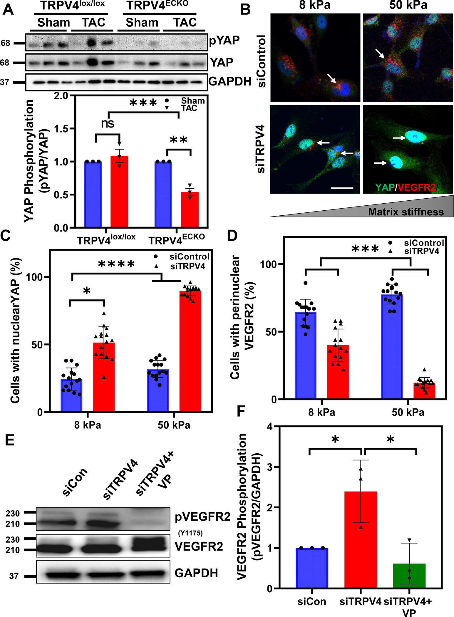
A) Representatives immunoblot and quantitative analysis showing decreased phosphorylation of YAP in TRPV4ECKO-TAC mice. Note molecular weight markers are shown in the left side B) Representative immunofluorescence images showing YAP (green) and VEGFR2 (red) in control and TRPV4 siRNA transfected human endothelial cells cultured on ECM gels mimicking normal heart stiffness (8kPa) and failing heart stiffness (50kPa). Note that perinuclear localization of VEGFR2 (red; arrows) in control siRNA-treated cells which is disappeared in TRPV4 siRNA-treated cells with concomitant nuclear translocation of YAP (green; arrows). C-D) Quantitative analysis showing the average number of cells/field (10–15 fields with around 200–300 total cells per condition from three independent experiments) with nuclear translocation of YAP and perinuclear VEGFR2. Representative western blots (E) and quantitative analysis (F) of phospho-VEGFR2 in TRPV4 siRNA downregulated EC in the presence or absence of YAP inhibitor, verteporfin (VP). Note molecular weight markers are shown in the left side; (n=3; Two-way ANOVA followed by Tukey post hoc analysis and Significance was set at *p<0.05; ***p<0.001 ****p<0.0001).