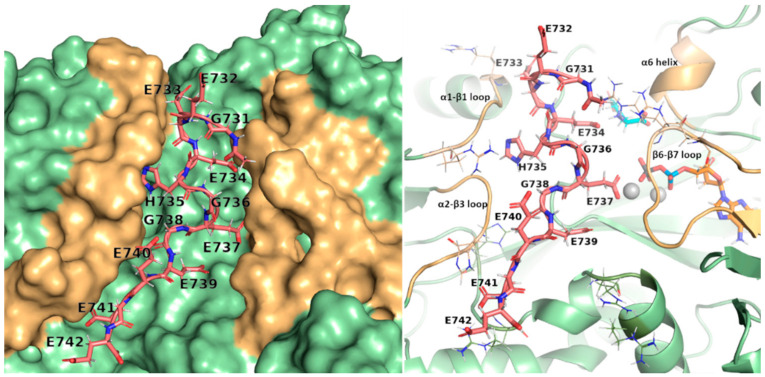
Figure 3.
Surface and cartoon view of TTLL5 active site. The peptide (in pink, shown as sticks) fits into the crevice centered at the active site, with Glu737 directed towards ADP. Glutamates align to form electrostatic interactions with the positively charged residues (shown as lines in the cartoon view), stabilizing the peptide into the pocket. The structural components surrounding the peptide chain are shown in light orange (loops α1-β1, α2-β3, β6-β7, and helix α6), with the rest of the protein shown in green; the donor glutamate is shown in cyan; magnesium ions and ADP are shown in gray and orange, respectively.

