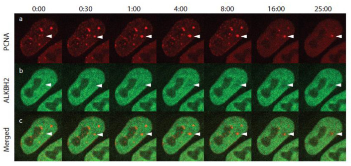
Figure 3.
Selected frames from a time-lapse movie of ALKBH2 and PCNA after UV micro-irradiation. (a) PCNA-mCherry; (b) ALKBH2-EGFP; (c) merged. White arrowheads indicate the micro-IR site. The shown S-phase cell exhibited a number of PCNA replication foci. Following micro-IR, these foci dissolved due to checkpoint activation.

