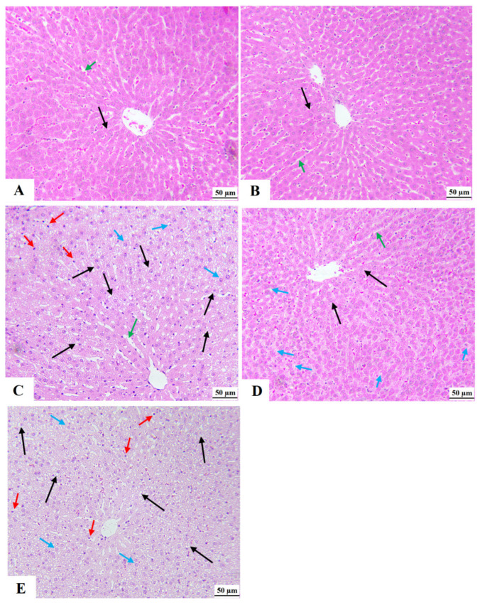
Figure 6.
Morphological images for the livers of all groups of rats as stained by hematoxylin and eosin stain (magnification = 200×). (A,B): were taken from control rat and XH-treated rats, respectively, and showed normal histological features, including central vein, hepatocyte (black arrows), and sinusoids (green arrows). (C): Represents HFD-fed rats and showed a massive increase in the number of cytoplasmic fat granules/vacuoles in the majority of the hepatocytes (black arrows). These livers also showed dilated sinusoids (green arrows) and an increased number of infiltrating immune cells (red arrows) and pyknotic cells (blue arrows). (D): was taken from HFD + XH-treated rats and showed much improvement in the structure of hepatocytes (black arrow) and normally sized- sinusoids. However, fat vacuoles were still seen in the hepatocytes that are away from the central vein but in smaller quantities as compared to HFD-fed rats in (C). (E): was taken from HFD + XH + CC and showed almost similar pathological changes to those seen in the HFD-fed rats.

