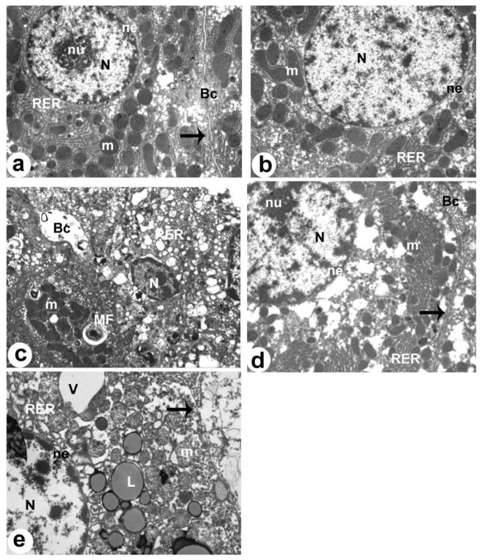
Figure 7.
Electron micrographs of the livers of all groups of rats as taken by electron microscopy. (a,b): were taken from control and XH-treated rats. Both livers showed a normal hepatocyte (H) with an intact nucleus (N), rough endoplasmic reticulum (RER), and mitochondria (m). The normally appeared nucleus was surrounded by an intact nuclear envelope (NE) and contained nucleolus (nu) and clear chromatin masses (Chr). The hepatocyte plasma membrane (arrow) was also seen and intact. Normal bile canaliculus (Bc) with intact microvilli were also seen. (c): was taken from NAFLD-treated rats and showed obvious apoptotic and degenerated hepatocytes with pyknotic nucleus (N), increased amounts of lipids (L), and myelin figures (MF). The RER was damaged and dilated. Dilated bile canaliculus (Bc) with damaged microvilli were also seen. (d): was taken from an HFD + XH-treated rat and showed improvement in the structure of the hepatocyte (H) and bile canaliculus (Bc) with partial changes in some mitochondria (m) and the RER. (e): was taken from HFD + XH + CC-treated rats and showed similar ultrastructural changes to those that appeared in the livers of HFD-fed rats with larger lipid droplets (L) and vacuoles (V).

