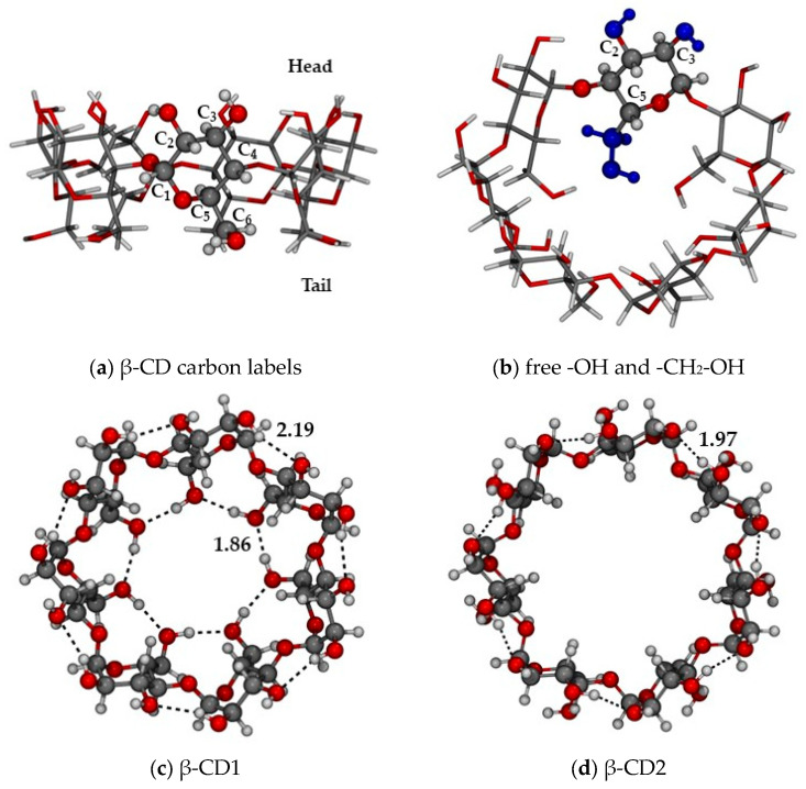
Figure 1.
(a) Carbon labeling of the α-glucopyranose ring and “Head”/”Tail” parts of β-cyclodextrin; (b) hydroxyl and hydroxymethyl groups free to rotate during the conformational search (highlighted in blue); (c) closed (CD1) and (d) open (CD2) conformations, as optimized at the r2SCAN-3c level. Color code: H in white, C in grey, O in red, H-bond as red dotted lines. H-bond distances in Å.

