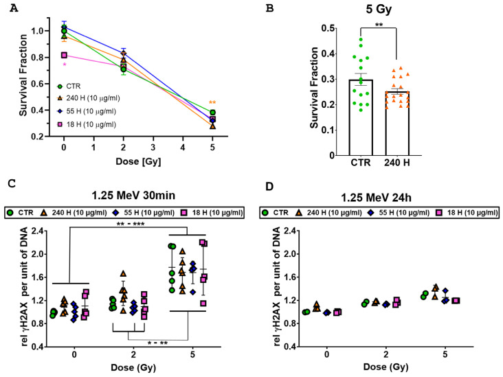
Figure 6.
Effect of combined H-ND and 1.25 MeV γ-ray treatment on DAOY cells. (A) Clonogenic survival assays’ mean survival relative to parental untreated cells and standard error of the mean are shown. All experiments were performed on at least 10 replicates. (B) Histogram plot highlighting the differences between cells irradiated with 5 Gy pre-treated with 240 nm H-NDs (orange triangles) or untreated (CTR, green dots). (C) γ-H2AX flow cytometry assay at 30 min after irradiation. (D) γ-H2AX flow cytometry assay at 24 h after irradiation. Data were represented as the mean florescence intensity (MFI) per unit of DNA normalized on the sham-irradiated untreated controls. Two-way ANOVA tests with the Greenhouse–Geisser correction and Tukey’s multiple comparison tests have been performed. The p-values ≤ 0.05 (*) and ≤0.01 (**) were considered as statistically significant; the p-values ≤ 0.001 (***) was considered as highly statistically significant.

