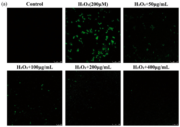
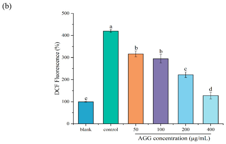
Figure 4.
Effects of the extracts on intracellular ROS in LPS-induced RAW 264.7 macrophages. (a) Assessment of ROS levels by confocal laser scanning microscopy with DCFH-DA fluorescent dye. ROS levels were evaluated by confocal laser scanning microscopy with DCFH-DA fluorescent dye. Green: DCF fluorescent representation of intracellular ROS. Scale: 100 μm. (b) Quantitative analysis of DCF fluorescence intensity percentage in bar graphs. Different letters (a–e) mean significantly different at p < 0.05. Note: control: without-H2O2 treatment group; blank: group treated with only 200 μM H2O2.


