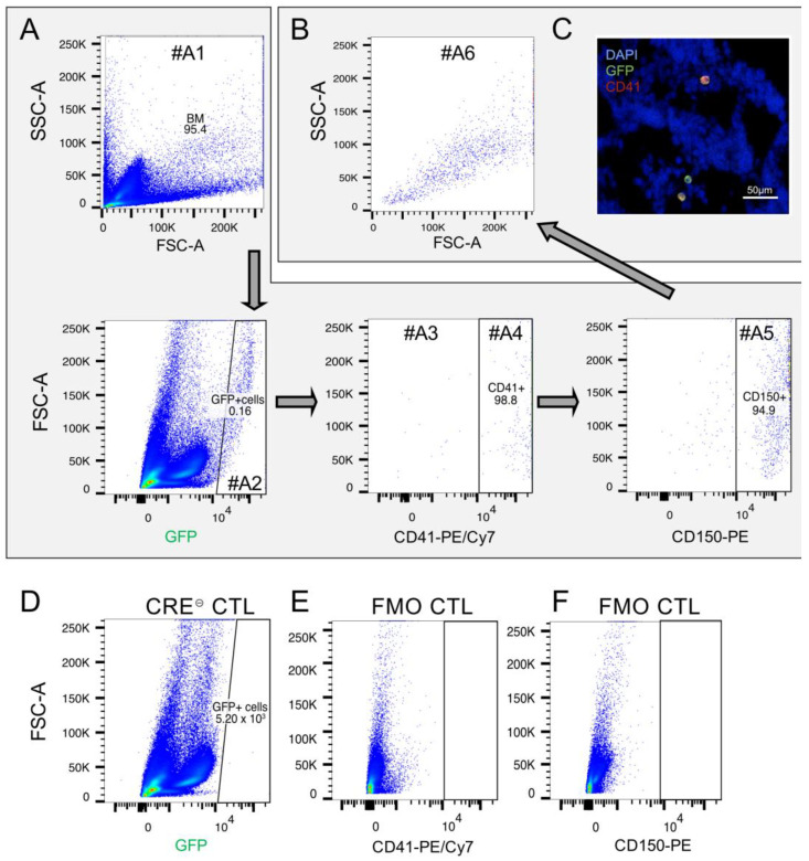
Figure 3.
Analysis of the PDGFb transgene-expressing cells in the spleen. (A) To detect the expression pattern of the PDGFb construct in spleen cells, PDGFb transgene reporter (GFP) was detected using FACS analysis on spleen samples. GFP-positive cells (gate #A2) were mainly CD41+/CD150+ (gate #A4, #A5) showing (B) big cell volume and high granularity (gate #A6) indicating megakaryocytic lineage properties. There was the rest of unidentified GFP+/CD41- cells, which were detected by FACS (gate #A3) as well as by (C) immunofluorescent staining on cryosections. Blue, DAPI; green, GFP; red, CD41. (D) Cre⊝ CTL spleen cells stained for GFP. (E) Fluorescence minus one (FMO) control for CD41-PE/Cy7. (F) Fluorescence minus one (FMO) control for CD150-PE.

