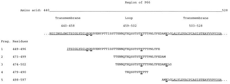
FIG. 2.
Monoclonal antibody H1337 epitope mapping. Shown are sequences of overlapping recombinant peptides (fragments 1 to 5) representing amino acids 440 to 528 of P66 of B. burgdorferi. Origins of fragments (Frag.) 1 and 5 of P66 and their Western blot reactivities with serum specimens from patients with Lyme disease have been described elsewhere (11). The amino acids of predicted transmembrane regions flanking the putative surface-exposed loop of P66 are underlined. Lysine and arginine residues at predicted trypsin cleavage sites are indicated by double underlines. Amino acids are numbered according to the processed P66 sequence of B. burgdorferi B31 (10).

