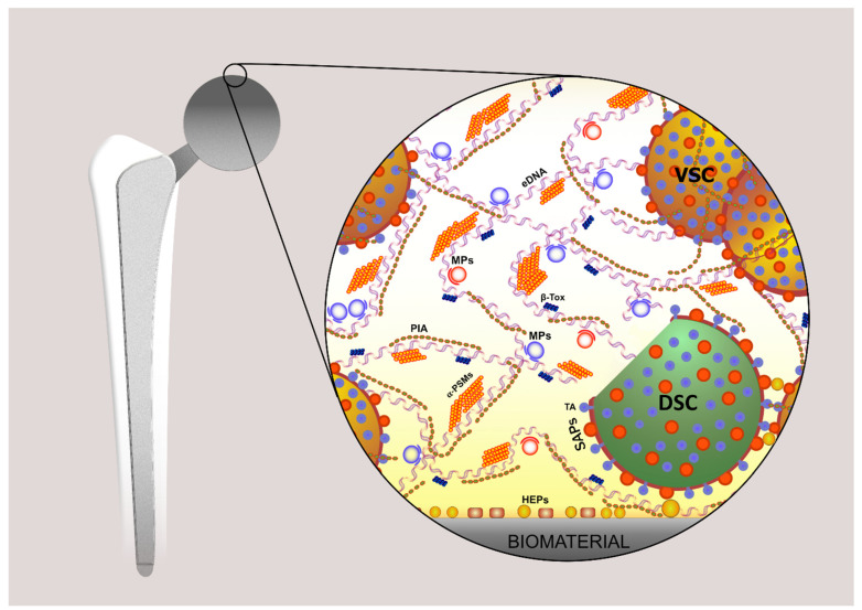
Figure 1.
This figure schematically illustrates the complex architecture of a biofilm of S. aureus established on the surface of and around a hip prosthesis. It shows, on the left, an infected hip prosthesis and, on the right, a magnified schematic view of the biofilm, revealing its main polymeric components: PIA, an exopolysaccharide, namely the polysaccharide intercellular adhesin; eDNA, extracellular DNA; SAPs, surface-associated proteins (e.g., SasG, clumping factor B, SdrC, Bap, FnBPA, and FnBPB); amyloidogenic proteins (e.g., β-Tox, β-Toxin; PSMs, phenol-soluble modulins such as α-PSM1); MPs, moonlighting proteins (cytoplasmic proteins with a moonlighting role in the biofilm matrix); HEPs, host extracellular matrix proteins; TA, teichoic acids; VSC, viable staphylococcal cell; DSC, dead staphylococcal cell. It must be taken into consideration that, under real in vivo conditions, a further level of complexity is associated with the existence of polymeric substances contributed by the host tissues themselves.

