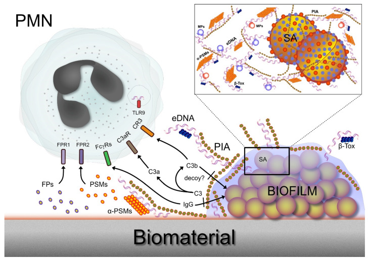
Figure 2.
This figure illustrates a polymorphonuclear neutrophil (PMN) that encounters an S. aureus biofilm on a biomaterial surface. A series of receptors that are expressed either on the surface or on the membranes of intracellular vesicles enable the PMN to directly recognize specific polymers taking part in the biofilm architecture or indirectly sense complement components generated following the triggering of the complement cascade by biofilm EPS. At the same time, EPS components such as PIA appear to protect bacteria from opsonization and their consequent targeting and phagocytosis by PMNs, as discussed in the next sections. The inset box on the upper right corner shows a magnified schematic view of the biofilm, revealing the complex weaving of extracellular polymeric components of matrix. Legend: amyloidogenic proteins (e.g., β-Tox, β-Toxin; α-PSMs, phenol-soluble modulins such as α-PSM1); C3, complement component 3; C3a, C3a protein formed by the cleavage of complement component 3; C3b, C3b protein formed by the cleavage of complement component 3; C3aR, complement component 3a receptor; C3R, complement receptor 3 (alternatively termed CD11b/CD18); eDNA, extracellular DNA; FPs, formyl peptides; FPR1 and FPR2, respectively, formyl peptides receptor 1 and 2 [20]; FcγRs, Fc receptors for IgG; MPs, moonlighting proteins (cytoplasmic proteins with a moonlighting role in the biofilm matrix); PIA, polysaccharide intercellular adhesin; PSMs, phenol-soluble modulins; SA, S. aureus cell; TLR9, Toll-like receptor 9 (expressed in intracellular vesicles).

