Figure 1:

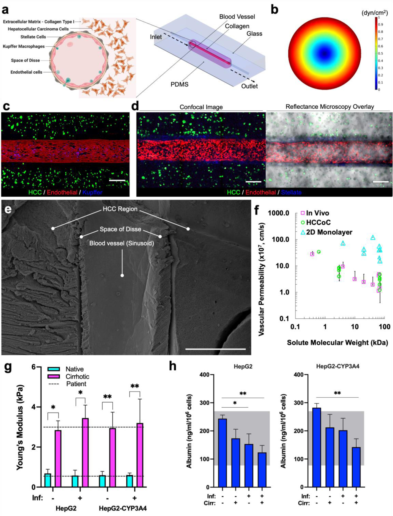
Vascularized HCCoC. a) CAD model of the microfluidic device and anatomical illustration of a liver sinusoid cross-section. The device consists of a single inlet and outlet with four major cell lines in liver. b) Finite element modeling of shear stress profile in HCCoC showing target 1 dyn/cm2 wall shear stress has been reached in the device. c) Device contains HCC (GFP), endothelial (mKate), Kupffer cells derived from THP-1 monocytes (Cell Tracker), and unlabeled stellate cells. d) Labeled stellate cells (Cells Tracker) in the space of Disse surrounds the circular blood vessel. e) SEM image of the HCCoC cross-section at the center of the blood vessel. f) Vascular permeability of vascularized HCC-on-a-chip, in vivo, and 2D monolayer findings in the literature at different solute sizes. [36–43] g) Compression modulus of HCCoC under normal and inflamed conditions tuned by collagen content to match native and cirrhotic stiffnesses and their comparison with HCC patient biopsy tumor reported in the literature. [44,45] h) Albumin secretion of HCC cells and comparison with reported HCC patient values. [46,47] All data is obtained on day three upon the completion of the preconditioning protocol. Scale: 300 μm. *p < 0.05, **p < 0.01. n = 4. All data represent means ± SD. HCC: hepatocellular carcinoma. CAD: Computer-aided design. HCCoC: Hepatocellular carcinoma-on-a-chip, GFP: green fluorescent protein. Inf.: Inflammation, Cirr: Cirrhosis.
