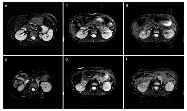
Figure 3.
Example of a case with residual tumor and local progression in a 10-year-old girl with ganglioneuroblastoma, N-MYC non-amplified tumor, INSS Stage IV. The T2 weighted images are shown. (A,B) Before resection, (C,D) after resection, and E and F during follow-up. (A,C,E) Transversal images at the level of the primary tumor on the left suprarenal side. After surgery and in the follow-up, no tumor can be detected (C,E). Enlarged left lymph node before surgery (B) and after surgery (D) (arrows). According to the surgical report, a CME was performed. In the course of 12 months, the tumor progressed (F) (arrow).

