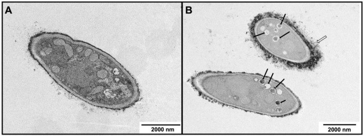
Figure 5.
Transmission electron microscopic (TEM) observations of the morphology of M. canis hyphae following exposure to ABT. (A) Control, in the absence of ABT and (B) with 500 μg/mL ABT. Open arrow indicates electrodense material at the cell wall surface and black arrows indicate vacuoles with debris.

