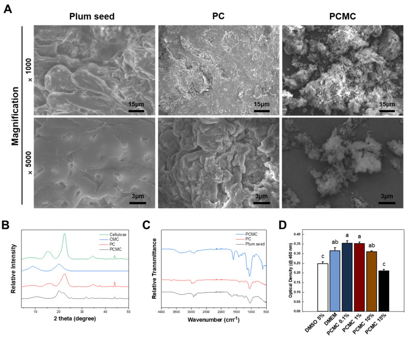
Figure 2.
Characterization of the PC and PCMC. (A) SEM image displaying the surface of the plum seed, PC, and PCMC. In plum seeds, distinctive pores are observed, and fibers are densely packed. While cellulose does not exhibit these pores, the fibers are still tightly packed. In PCMC, the fibers are dispersed widely and do not exhibit a clustered configuration. (B) Crystalline structure of cellulose by X-ray diffraction. The peaks of PC and PCMC appeared to be similar to those of commercial cellulose and CMC, respectively. (C) FT-IR spectra of PC and PCMC. These peaks confirm that PC shares similarities with common cellulose in terms of chemical structure. (D) WST-1 assay according to the concentration gradient of PCMC. Cytotoxicity evaluations were conducted at various PCMC concentrations to determine the maximum tolerable concentration before adjusting the PCMC ratio as bioink. Error bars indicate the standard mean of errors. The same letter means statistical insignificance (Duncan’s new multiple range test, p ≤ 0.05). SEM, scanning electron microscopy; FT-IR, Fourier transform infrared; WST, water soluble tetrazolium.

