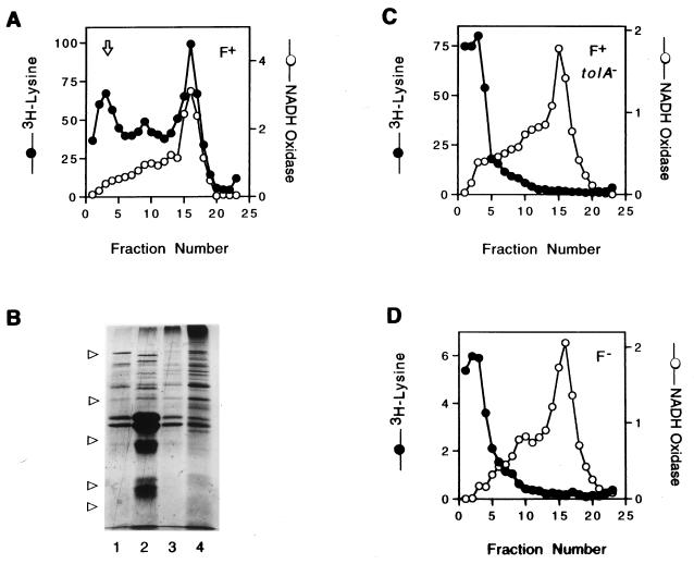
FIG. 1.
Phage coat protein pVIII from infecting phages is not found in the inner membranes of either F− or tolA mutant bacteria. (A, C, and D) Cultures of K17DE3 bacteria that were F+ (A), tolA/F+ (C), or F− (D) were infected with [3H]lysine-labeled phages (Table 1, experiment 1). The washed bacteria were broken in a French press, and the membrane fractions were separated by sucrose flotation gradient as described in Materials and Methods. The fractions, collected from the bottom of the gradient, were assayed for NADH oxidase activity, and radioactivity, which is expressed as counts per minute (in thousands), was determined. The arrow in panel A indicates the flotation position of both intact and broken phages. (B) Coomassie blue-stained SDS polyacrylamide gel of pooled fractions 1 to 5 (lane 1), 7 to 10 (lane 2), 11 to 13 (lane 3), and 14 to 18 (lane 4) from panel A. The arrows on the left indicate migration of protein standards with molecular masses (from the top) of 97, 45, 31, 21.5, and 14.4 kDa.

