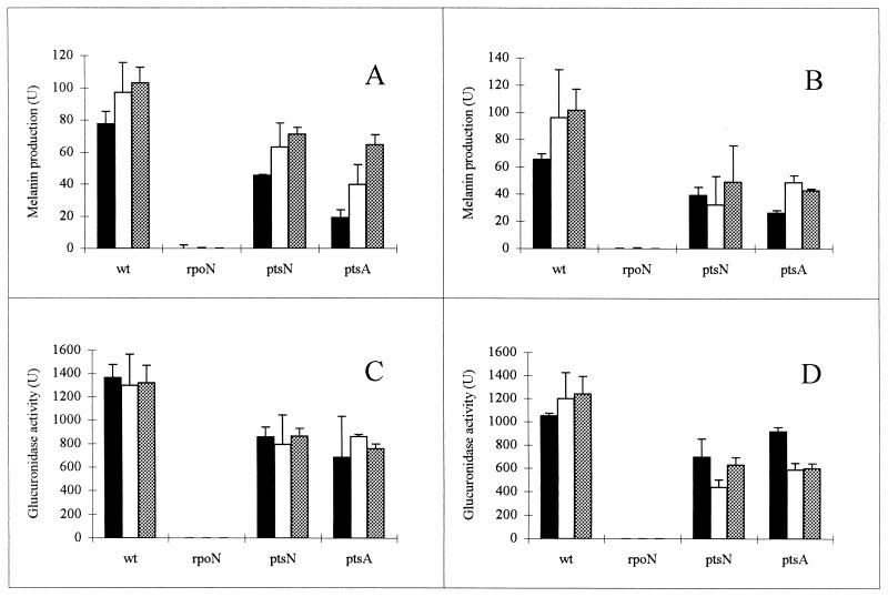
FIG. 3.
Melanin production (A and B) and expression of nifH (C and D) in wild-type R. etli (wt) and rpoN (FAJ1154), ptsN (FAJ1165), and ptsA (FAJ1166) mutants. All data are the means from four independent replicates. Error bars denote the standard deviations. Precultures were grown overnight in TY medium at 30°C, diluted 20-fold in the different media, and incubated overnight with 0.5% oxygen (34). The nitrogen source used was alanine (20 mM). The carbon sources were mannitol (black bars), succinate (stippled bars), and malate (white bars) at 5 mM (A and C) and 20 mM (B and D). To quantify melanin production, cultures were lysed at 37°C in the presence of a solution containing sodium dodecyl sulfate (1%), CuSO4 (10 μg/ml), and tyrosine (30 μg/ml). The OD of the culture after lysis was measured at 340 nm in a microplate reader after 60 and 120 min of incubation. The difference between the ODs at 340 nm was used to calculate the units. Units are expressed as the ratio of the change in OD at 340 nm to the OD at 595 nm. β-Glucuronidase activities of the translational pnifH-gusA fusion plasmid pFAJ21 are expressed as Miller units.

