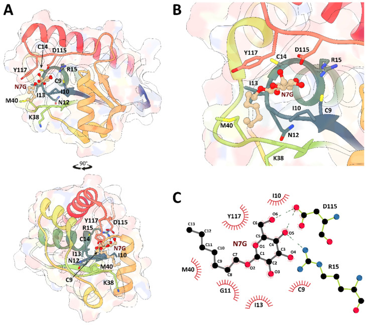
Figure 3.
(A) Cartoon and molecular surface representation of EaAmsI bound to N7G as a result of the DOCK6 molecular docking. EaAmsI cartoons are colored from blue to red going from the N- to the C-terminal. Residues involved in the reaction or in the binding of N7G are sticks, while N7G is in ball-and-stick (carbon, light orange; oxygen, red). (B) Detail of N7G binding pose in the EaAmsI binding site. (C) Scheme of the interactions between the docked N7G molecule and EaAmsI. The scheme represents H-bonds (dashed lines) and van der Waals interactions (red spiked arcs).

