Figure 2.

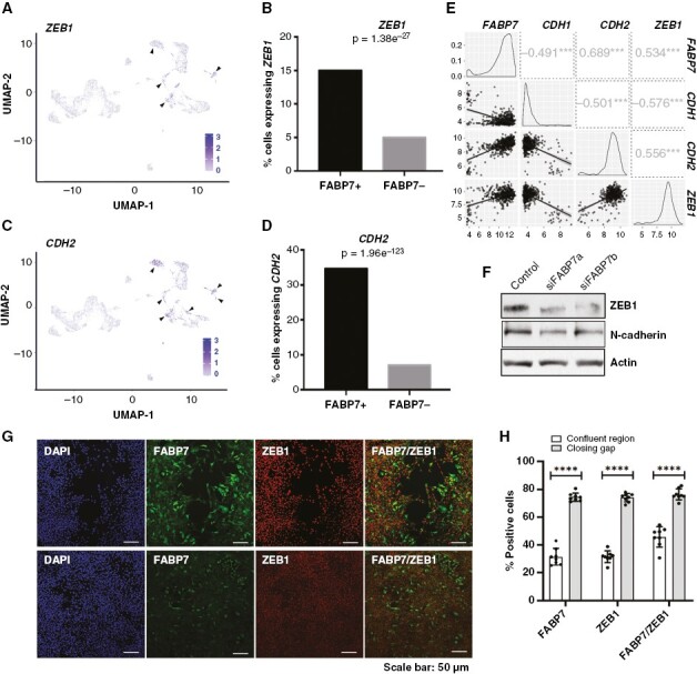
Expression of epithelial-to-mesenchymal transition markers is correlated with and regulated by fatty acid-binding protein 7 (FABP7) in GBM cells. (A, C) UMAP depicting ZEB1 (A) and CDH2 (C) expressing cells in GBM cell clusters generated through scRNA-seq analysis. (B, D) Analysis of the scRNA-seq data showing significantly higher percentages of cells expressing ZEB1 (B) and CDH2 (encoding N-cadherin) (D) in FABP7-positive cells compared to FABP7-negative cells. (E) Correlation of FABP7 RNA levels with that of ZEB1 and CDH2 in a TCGA dataset (HG U-133A) comprised of 453 GBM patients. Numbers in squares denote correlation coefficients, ***, P < .001. (F) Western blot showing decreased expression of ZEB1 and N-cadherin resulting from FABP7 knockdown in A4-004 cells cultured in neurosphere medium. (G) Representative images showing coimmunostaining of FABP7 and ZEB1 (G) in A4-004 GSCs in the closing gap and confluent regions 24 hours after introduction of the scratch. (H) Histogram showing quantitation and significance test of the data from the FABP7, ZEB1 and combined FABP7/ZEB1 fluorescence immunostaining data shown in (G). N = 2, in triplicate. **** denotes P < .0001.
