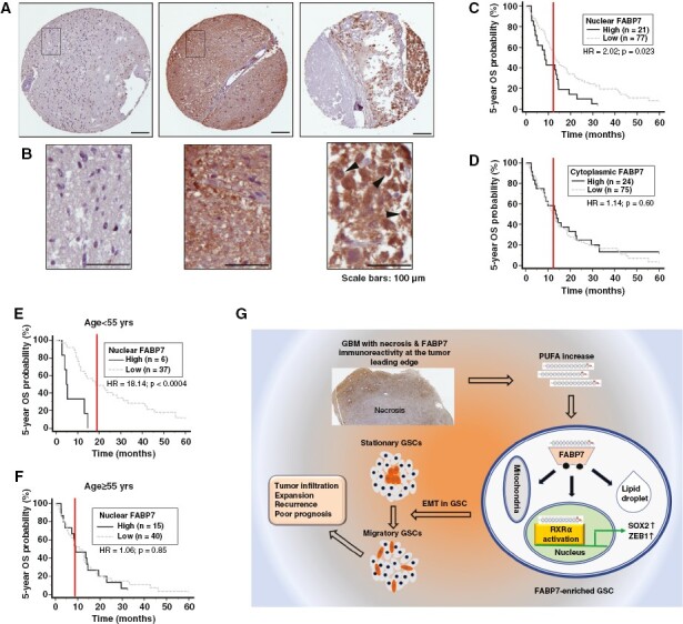
Figure 6.

Immunohistochemical analysis of a GBM TMA. (A) Representative immunostaining images showing negative (left panel), uniform strong cytoplasmic and nuclear (middle panel), and heterogenous intratumoral and subcellular distribution of Fatty acid-binding protein 7 (FABP7) (right panel). (B) Magnified images from the squared regions shown in (A). Arrowheads point to FABP7 immunoreactivity in cell nucleus. (C, D) Patient overall survival (OS) curves generated based on FABP7 nuclear (C) and cytoplasmic immunoreactivity (D). (E, F) Patient overall survival curves generated based on nuclear FABP7 immunoreactivity in a patient population under (E) or above (F) the median age of 55. Three to six cores from each tumor on TMA slides were immunostained and scored. HR denotes hazard ratio. Scale bars: 100 μm. (G) Graphical summary of the role of the FABP7-RXRα neurogenic pathway in determining stationary to migratory transition in GSC and tumor progression.
