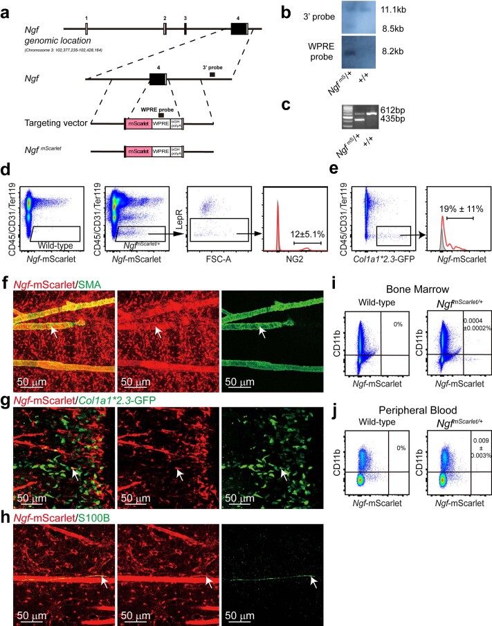
Extended Data Fig. 1. Generation and characterization of the NgfmScarlet mouse reporter allele.
(a-c) The mouse Ngf gene was modified by inserting an mScarlet-WPRE-pA cassette after an alternative ATG start codon in exon 4, replacing most of the coding sequence in exon 4. Open boxes indicate untranslated regions and black boxes indicate translated regions of Ngf. The correctly targeted founder mouse (F0) was identified by southern blotting (b) using 3’ and WPRE probes (black bars in a) (representative of three independent experiments). (c) PCR genotyping of genomic DNA confirmed germline transmission of the Ngf mScarlet allele. Mice were backcrossed at least three times onto a C57BL/Ka background before analysis (representative of three independent experiments). (d) Flow cytometric analysis showed that 12% of Ngf-mScarlet+ bone marrow stromal cells were NG2+ smooth muscle cells in enzymatically dissociated bone marrow cells. (e) When bones were crushed and enzymatically dissociated, 19% of osteoblasts were Ngf-mScarlet+ (4 mice from 4 independent experiments). (f-h). Deep imaging of femur bone marrow from 2-8 month-old Ngf mScarlet/+ mice. The Ngf-mScarlet+ cells included SMA+ periarteriolar smooth muscle cells (arrow, f), a subset of Col1a1-GFP+ osteoblasts associated with trabecular bone in the metaphysis (arrow, g), and S100+ Schwann cells associated with nerve fibers in the bone marrow (arrow, h) (each panel reflects data from three mice from three independent experiments). As in Fig. 1, most of the Ngf-mScarlet+ cells in these images were LepR+ perisinusoidal stromal cells. (i, j) Flow cytometric analysis showed that only rare macrophages in the bone marrow (i) or blood (j) were Ngf-mScarlet+ (3 mice from 3 independent experiments).

