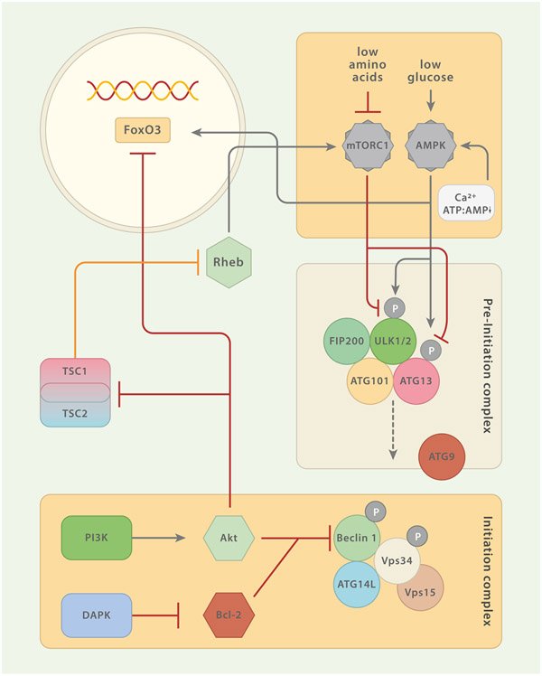
Fig. 2.
Regulatory Circuits of Macroautophagy (MA). The pre-initiation and the initiation complexes constitute the most proximal modules of MA. Several constituents of these complexes are under the control of regulatory elements. Both, AMPK and mTORC1 sense nutrients and are the two major regulatory units that control MA. mTORC1 inhibits MA by phosphorylation of pre-initiation complex member ULK1/2 (and/or ATG13). mTORC1 activity is promoted by amino acid sensing Rag GTPases which also mediate the translocation of mTORC1 to the lysosomal membrane. This translocation is important for the mTORC1-promoting activity of Ras homolog enriched in brain (Rheb). The tuberous sclerosis complex (TSC) 1/TSC2 can enhance MA by repressing Rheb. Akt reduces macroautophagic activity by inhibiting TSC1/TSC2 or via interaction with the transcription factor FoxO3. Moreover, growth factors may signal through phosphatidylinositide 3-kinase (PI3K) to Akt, which in turn inhibits the Beclin 1-containing class III PI3K complex. The calcium/calmodulin (Ca2+/CaM) serine/threonine kinase death-associated protein kinase (DAPK) activates MA via phosphorylation of Beclin 1 (initiation complex) which entails dissociation of Beclin 1 from Bcl-2. AMPK is a central positive regulator of MA. Phosphorylation of ULK1/2 (and/or ATG13) at residues different from those targeted by mTORC1 and direct interaction with FoxO3 augment MA activity. AMPK itself can be triggered by increasing levels of AMP relative to ATP and free Ca2+.

