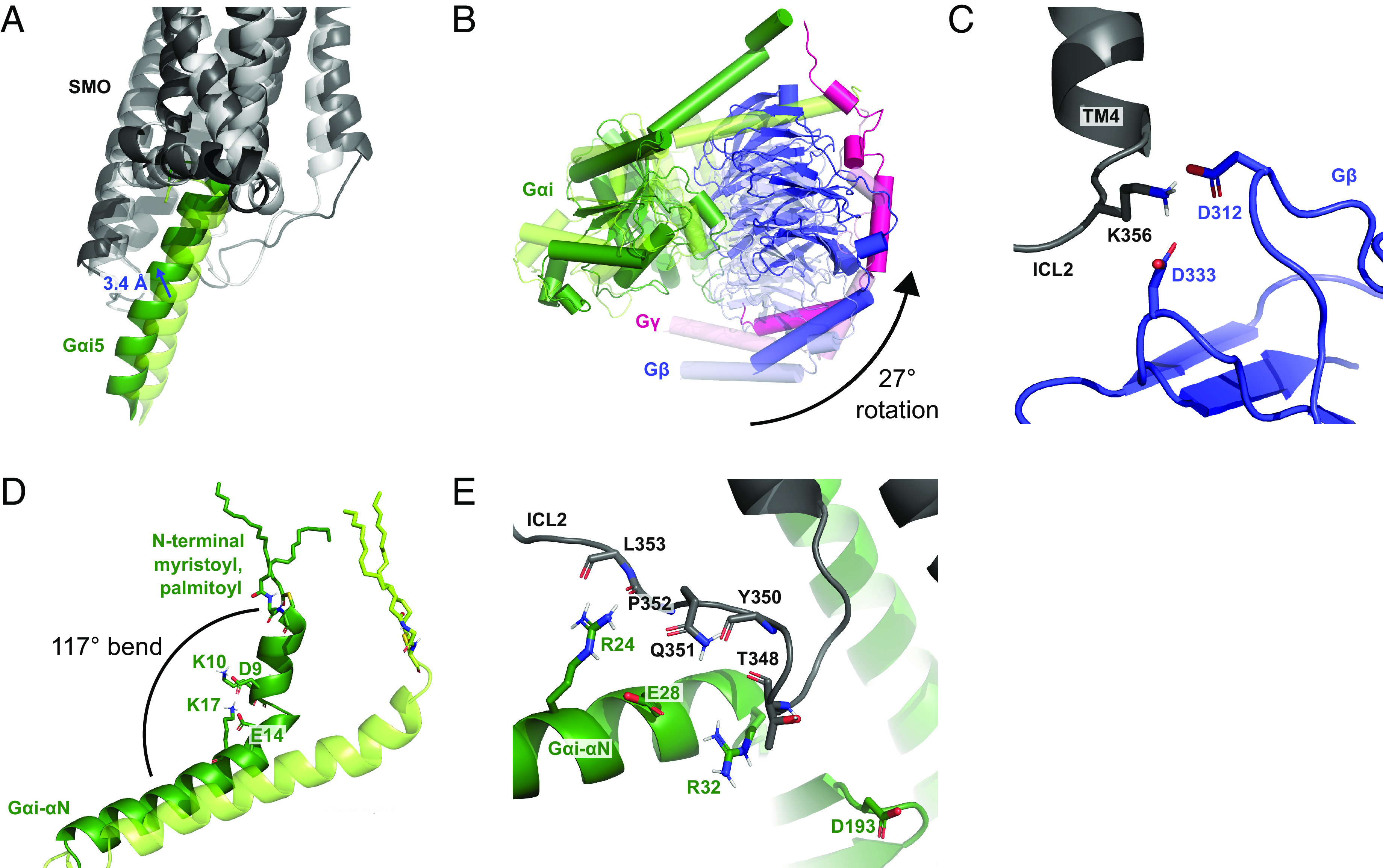
Fig. 2.

Reorientation of the heterotrimeric Gi relative to SMO over a 370-ns MD simulation. (A) Comparison of the Gαi5 binding position in the initial (0 ns, light tint) and final structure (370 ns, dark tint). (B) Comparison of the Gi protein subunits between the initial (0 ns, light tint) and final structure (370 ns, dark tint). (C) Interaction between ICL2 and Gβ at 370 ns. (D) Movement of the Gαi-αN helix toward the membrane between the initial (0 ns, light tint) and final structure (370 ns, dark tint). (E) Interaction between ICL2 and Gαi at 370 ns.
