
Fig. 3.
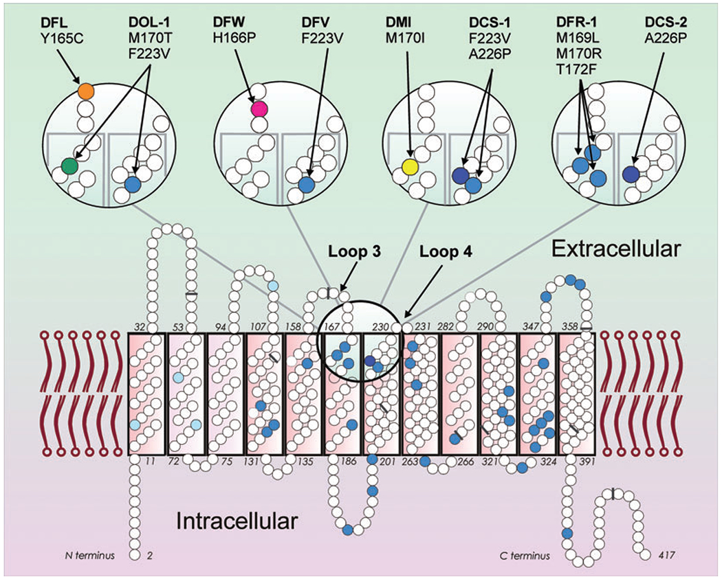
Partial D with amino acid substitutions at the extracellular RhD protein vestibule. The two-dimensional model of the RhD protein (bottom) with 417 amino acids (small circles) depicts amino acids that differ in RhCE (blue) with the four C-typical substitutions (light blue) and the one E-typical substitution A226P (dark blue). The vestibule is lined in part by amino acids of loops 3 and 4 (large circle). This region is shown in more detail (top). The amino acid substitutions characteristic of eight partial D are indicated (colored circles and arrows). Four partial D harbor RhCE-like substitutions (blue circles). The nine exon boundaries in the RHD cDNA, as reflected in the amino acid sequence, are indicated (gray bars).