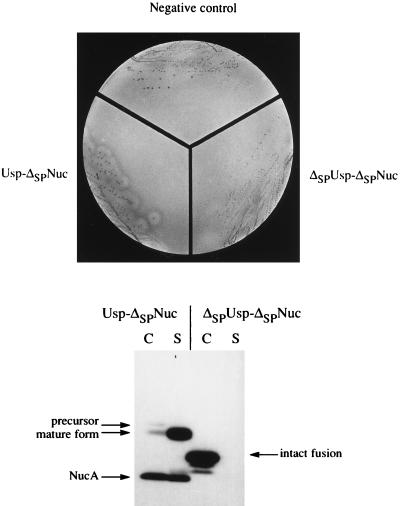
FIG. 2.
Specific detection of ΔSPNuc fusions which have an export signal in L. lactis. (Top) Nuc activity of Usp-ΔSPNuc but not of ΔSPUsp-ΔSPNuc is detected by plate tests. MG1363 strains containing plasmid pFUN (negative control), pVE8009 (Usp-ΔSPNuc), or pVE8010 (ΔSPUsp-ΔSPNuc) were streaked onto solid medium (brain heart infusion agar) and grown overnight. Colonies were overlaid with indicator medium containing toluidine blue, denatured DNA, and agar. Nuclease activity is detected by pink halos around colonies (Nuc+ phenotype) only in the case of MG1363(pVE8009), which produces Usp-ΔSPNuc. (Bottom) Location of Usp-ΔSPNuc and ΔSPUsp-ΔSPNuc fusion proteins in L. lactis. Cell (C) and supernatant (S) fractions of mid-exponential-phase cultures of L. lactis MG1363 derivative strains producing either Usp-ΔSPNuc or ΔSPUsp-ΔSPNuc fusion protein (from plasmids pVE8009 and pVE8010, respectively) were analyzed by SDS-PAGE and Western blotting using polyclonal Nuc antibodies. Putative forms of each fusion are indicated. Commercial NucA, used as a size reference (not presented), comigrates with the smallest degradation product of Usp-ΔSPNuc. Note that aberrant relative migration of Usp-ΔSPNuc forms (precursor, mature, and NucA) has previously been observed (30, 31) and that NucA in the cell fraction may be cytoplasmic or externally cell associated (31).

