
FIG. 3.
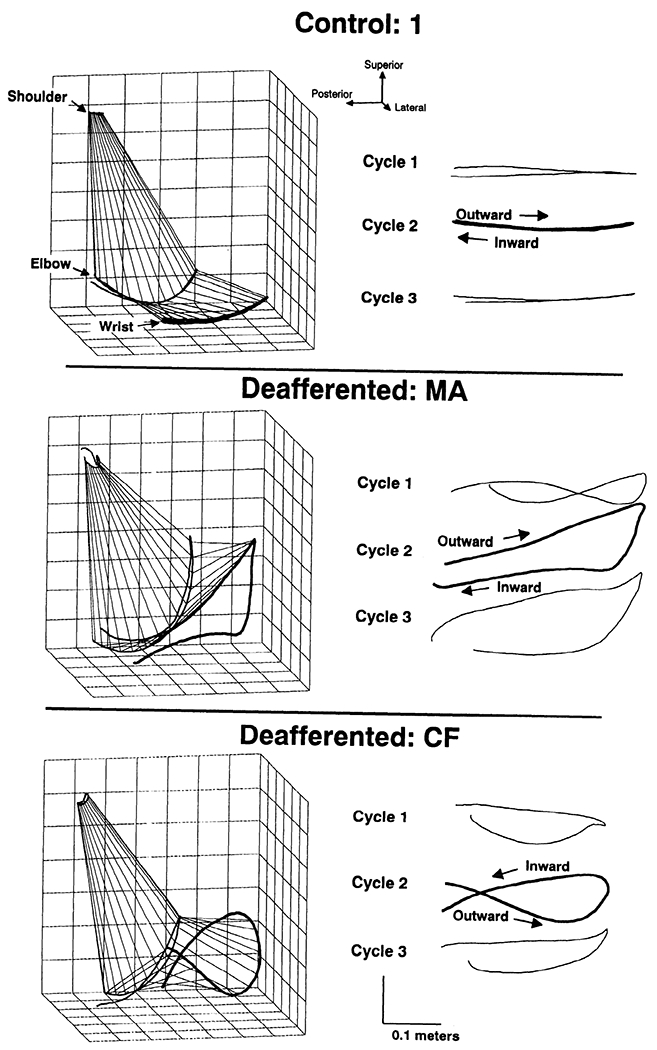
Left: 3-dimensional limb trajectories. A single cycle of a movement trial performed by control 1 (top) and the two leafferented patients: MA (middle) and CF (bottom). Shoulder, elbow (gray), and wrist paths are shown. Stick figures epresent limb positions at the beginning and end of each movement cycle. Calibration grid lines are spaced 0.05 m apart tight: parasagittal views of successive cycles of the wrist path. The second cycle of each movement (bold) corresponds to the 3-dimensional view shown on the left.