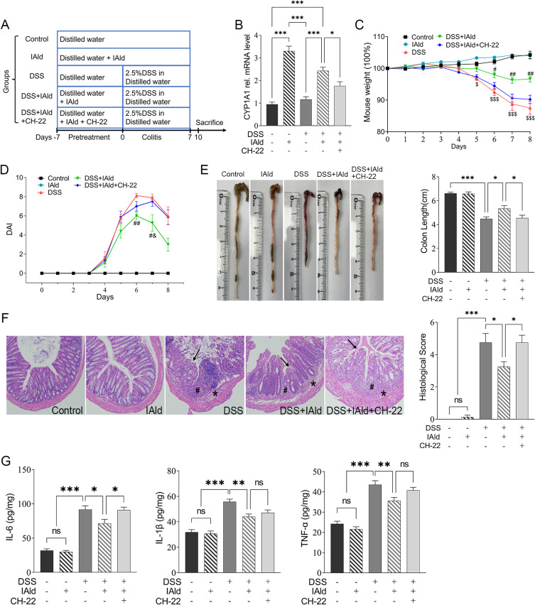
Figure 1.
Experimental schedule and effects of IAld supplementation on basic indicators in a DSS-induced colitis mouse model.
Notes: (A) Animal treatments schedule. (B) The mRNA expression of CYP1A1 in each group. (C) The measurements of bodyweight in each group. (D) The measurements of DAI scores in each group. (E) Representative images of colon length and quantitation of colon length of each group. (F) Representative images of haematoxylin and eosin staining of the colonic tissue and quantitation of histological score in each group. Arrows, ulceration; #, transmural inflammation; *, mucosal immune cell infiltration, magnification is X20 (G) The expression of IL-6, IL-1β and TNF-α in the colonic tissue were measured by enzyme-linked immunosorbent assay. The data are shown as the mean ± SEM (n = 8/group). * p < 0.05, ** p < 0.01, *** p < 0.001; # p < 0.05, ## p < 0.01 versus DSS; &p < 0.05 versus CH-22; $p< 0.05, $$$ p < 0.001 versus Control; ns, statistically non-significant.

