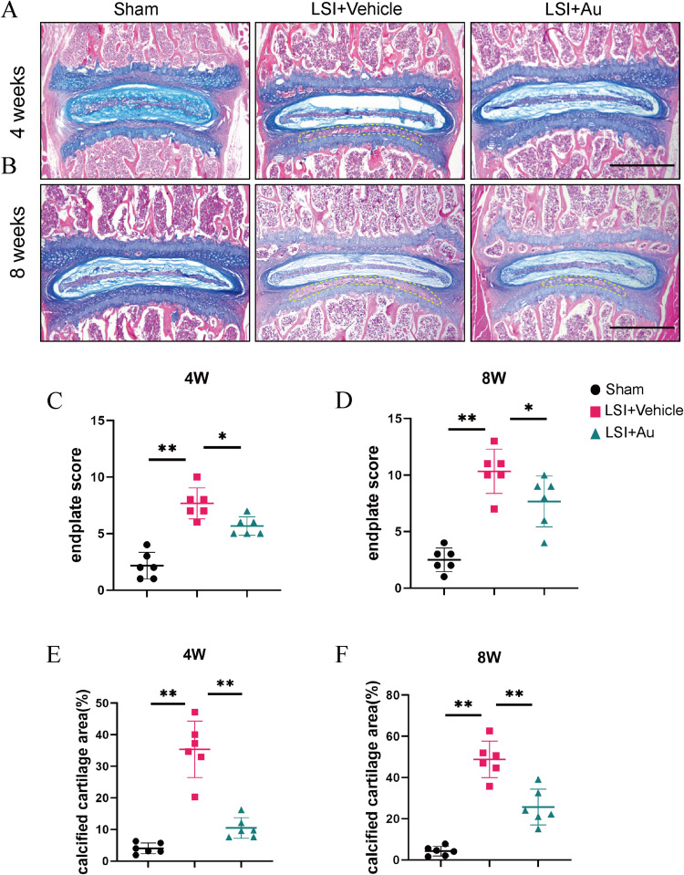
Figure 3.
Au attenuated cartilaginous endplate calcification in LSI mice. (A and B) Alcian Blue Hematoxylin/Orange G staining of cranial L4-5 endplate at 4 weeks and 8 weeks after operations. Yellow dashed boxes indicated regions shown as calcification. Scale bar = 500 μm. (C and D) Quantitative analysis of L4-5 endplate score at 4 and 8 weeks post-operation. (E and F) Quantitative analysis of L4-5 endplate calcified cartilage area at 4 and 8 weeks post-operation. The dose of Au was 10 mg/kg/day. Data were presented as means ± S.D. *p < 0.05; **p < 0.01; n = 6 in each group.

