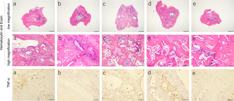
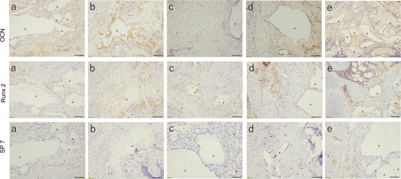
Fig. 9.
Histological analysis after 21 days of implantation in the femoral head: (a) CaP/CMC, (b) 3mixCaP, (c) CaP/PEI/pVEGF/SiO2, (d) CaP/PEI/pBMP-7/SiO2, and (e) CaP/PEI/siRNA- TNF-α/SiO2. The asterisk shows remaining implanted material (CaP). Scale bars: 1600 μm in low magnification of Hematoxylin and Eosin and 100 μm in high magnification of Hematoxylin and Eosin, TNF-α, OCN, Runx2 and SP-7.


