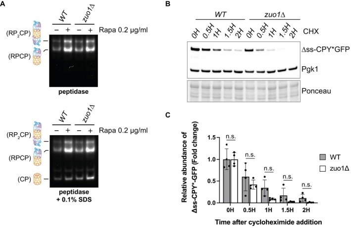
Figure 2. Proteasome homeostasis is not impaired in zuo1Δ cells.

- Yeast extracts from cells treated with 200 nM rapamycin for 3 h or left untreated were separated by native‐PAGE (3.8–5% gradient) and peptidase activity detected using the fluorogenic substrate Suc‐LLVY‐AMC in the presence or absence of 0.1% SDS. RP2CP, double‐capped proteasome; RPCP, single‐capped proteasome; and CP, core particle complexes are indicated.
- Immunoblot analysis of lysates from WT and zuo1Δ cells expressing Δss‐CPY*GFP from a plasmid treated with 35 μg/ml cycloheximide for the indicated time. Ponceau and Pgk1 staining served as the loading control.
- Graph shows densitometry analysis (mean ± s.d.) of the relative abundance of Δss‐CPY*GFP (normalised to Pgk1 levels) from (D) relative to the 0H time point. Statistical significance was assessed using two‐way ANOVA t‐test (n = 4 independent biological replicates). n.s. (not significant).
Source data are available online for this figure.
