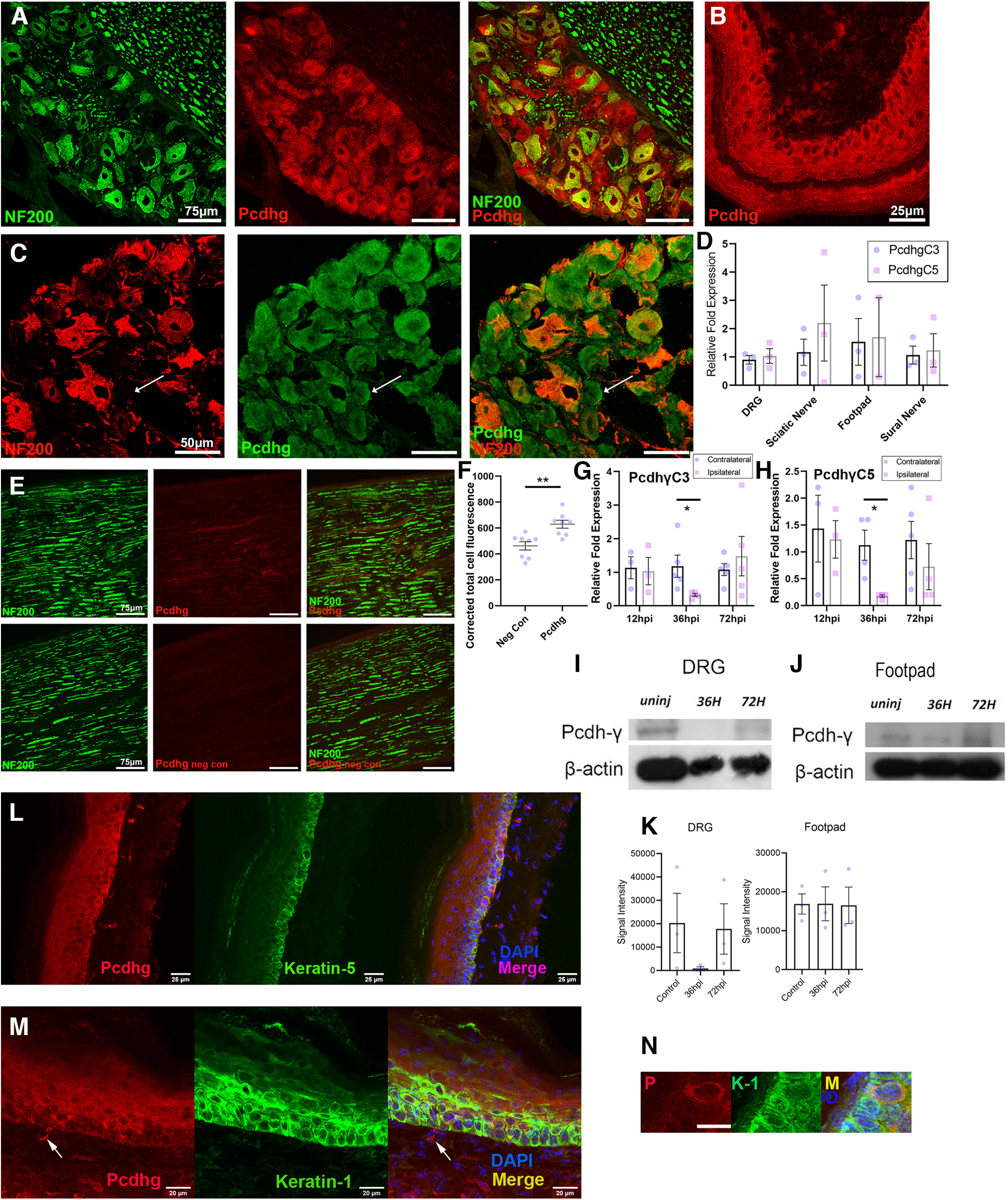
Figure 1.

Pcdhγ protein and mRNA is expressed in the mammalian peripheral nervous system. Transverse sections of mouse (A) and rat (C) DRGs immunostained for NF200 and Pcdhγ. Arrow indicates a smaller diameter neuron positive for Pcdhγ but negative for NF200. Pcdhγ is also expressed in keratinocytes in transverse sections of mouse footpads (B). DRG, sciatic nerve, footpad, and sural nerve were all positive for mRNA transcripts of the gC3 and gC5 exons (D). Sciatic nerve showed low intensity expression of Pcdhγ compared with a negative control and quantitated (E, F). (F; **p = 0.0022, two-tailed Student's t test, n = 8/group). PcdhgC3 (G) and gC5 (H) mRNA transcripts in DRGs show a significant drop in expression 36 h on the ipsilateral side postinjury but return to normal levels by 72 h [(G) PcdhγC3 *p = 0.02 contra vs ipsi PcdhgC3 36 h, two-tailed Mann–Whitney, n = 5 contra, 4 ipsi; (H) PcdhgC5 *p = 0.026 contra vs ipsi PcdhγC3 36 h, two-tailed Mann–Whitney, n = 4/group]. Representative tissue western immunoblots (I, J) with quantitation (K) from intact DRGs and footpads and ipsilateral to axotomy injury at 36 and 72 h (values not significantly different). DRG, dorsal root ganglion; NF200, neurofilament heavy subunit. Images of coexpression of keratin-5 (L) and keratin-1 (M, N, higher power) with Pcdhγ confirming Pcdhγ colocalization within keratinocytes (arrow in M shows an axon also staining). Scale bar = 25 μm for L, 20 μm for M, and 10 μm for N.
