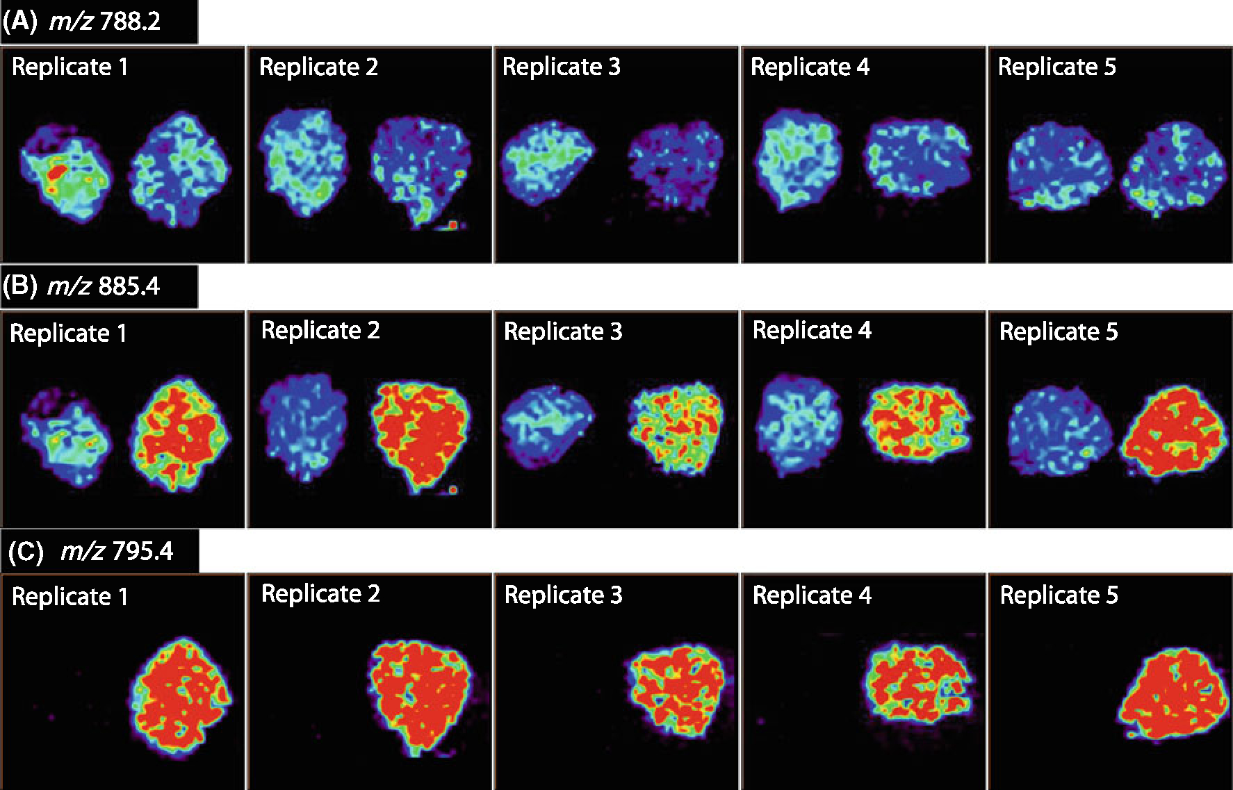
Figure 4.

Negative ion mode imaging of seminoma and adjacent normal tissue of five replicate samples of UH0303–05. (A) Ion images of m/z 788.2 PS(18:0/18:1), (B) ion images of m/z 885.4, PI(18:0/20:4), (C) ion images of m/z 795.4, seminolipid (16:0/16:0)
