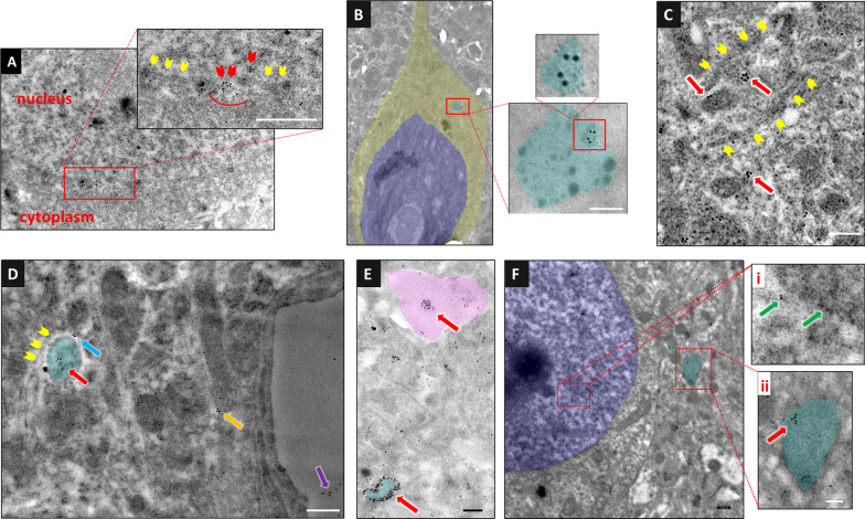
Fig. 2.
Immunoelectron microscopy illustrates that HMGB1 is released within small EVs. HMGB1 was labeled using 10 nm diameter gold-conjugated secondary antibodies on a perfusion-fixed brain section 15 min (A–E) and 3 h (F) after CSD. A The magnified image of a neuronal nucleus and cytoplasm in the inset shows gathering of gold particles (HMGB1) in areas where the nuclear membrane is not visualized (red arrowheads), suggesting that HMGB1 passes to the cytoplasm through nuclear pores, whereas no such accumulation is observed in the regions where the nuclear membrane is clearly visible (yellow arrowheads). B In another neuron, a multivesicular body (light blue) containing several sEVs, one of which harbors gold particles marking HMGB1, is seen. In this image, the focus was set on the gold particles to emphasize them at the expense of partly losing the details of cellular structures including the MVB membrane. However, the MVB can easily be identified by its more electrodense cytoplasm. C sEVs bearing HMGB1 molecules (red arrows) are seen within an astrocyte process (upper left) and the neighboring neuron soma (lower right). The plasma membranes of the neuron and astrocyte processes abutting each other (yellow arrowheads) are clearly visible. D The clustering of HMGB1 molecules (red arrow) presumably in an sEV inside an astrocyte process (light blue) abutting a neuron soma and in another sEV (blue arrow) between the neuron and astrocyte process are noticed. The plasma membrane of the neuron (yellow arrowheads) and the astrocyte process are visible. On the right, clustered HMGB1 molecules (possibly packed in a sEV) are also seen in an astrocyte end foot (orange arrow) and capillary lumen (purple arrow). E A sEV bearing clustered HMGB1 molecules (red arrow) within the tip of an astrocyte process (pink) and large numbers of gold particles lining the outline of an endosome (light blue) are easily visible. In this image, the focus was set on the gold particles as in panel B. F sEVs containing HMGB1 were still present in the cortex 3 h after CSD. Whereas gold-conjugated HMGB1-positive particles are scattered inside the nucleus of a neuron (i, green arrows), clustered gold particles (red arrow) are seen in a cytoplasmic vesicle (endosome, ii, light blue). The scale bars are 100 nm

