Fig. 3.
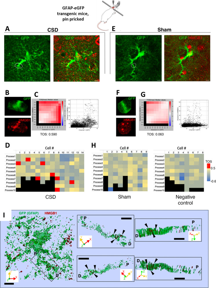
EVs loaded with HMGB1 are taken up by astrocyte processes. To assess the prevalence of HMGB1-loaded EV uptake by astrocytes, we examined the colocalization of HMGB1-immunopositive puncta with astrocyte processes visualized with anti-GFP antibodies in transgenic mice expressing GFAP-GFP and subjected to CSD. A and E show two representative cells from the CSD and Sham groups, with zoomed-in views of the outlined regions of interest (ROIs) (B and F). Scale bars: 10 µm. Scatter plots (C, G) show the distribution of corresponding pixel intensities in GFP and HMGB1 channels in the indicated ROIs. Metric matrices for threshold overlap scores (TOS) are shown in D, H with top percentages of highest intensity pixels in each ROI indicated in columns and rows for GFP and HMGB1, respectively. Each cell with color code in the matrix shows TOS values when the corresponding percentage of highest intensity pixels are selected in each channel, + 1.0 indicating absolute correlation, -1.0 indicating absolute anti-correlation, and 0 indicating random distribution and overlap between two channels. TOS values are not informative when one threshold is 100%; hence, the left column and bottom row are shown in black. Despite heterogeneity in the HMGB1 content of astrocyte processes and the presence of nonspecific punctate signals, the EzColocalization algorithm of FIJI disclosed that in the CSD cortex (n = 3 animals), 7 out of 14 astrocytes had at least one process with a TOS above 0.5, indicating a strong positive colocalization of HMGB1 and GFP signals, while none of the 17 astrocytes analyzed from sham (n = 2) and negative staining control animals (n = 2) was over that threshold, suggesting that uptake of released HMGB1-loaded EVs by astrocytes was not uncommon. I 3D surface reconstruction of a GFP-positive astrocyte and its process shows that HMGB1 immunopositive puncta (black triangles) are located inside the process but not superimposed falsely due to intensity projections. Black rectangle on the left panel indicates the HMGB1-immunopositive process that is visualized on the right panels from different angles in 3D. P and D indicate the proximal and distal ends of the process, respectively. Scale bars: 2 µm. X, Y, and Z axes of the volume are shown for orientation

