Fig. 4.
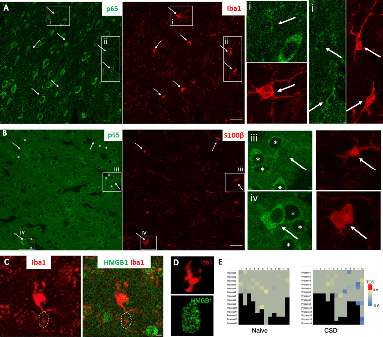
EVs loaded with HMGB1 are not observed in microglial processes. A No nuclear translocation of NF-ĸB p65 (green), a marker of proinflammatory transcriptional activity, was detected in microglia (Iba-1 immunopositive, red, arrows) 24 h after CSD. The round cell body and thin, long processes of microglia (arrow) also indicate that they are in an inactive state. Magnified images on the right of the boxed areas (i and ii, corresponding images in green and red) illustrate the presence of perinuclear (cytoplasmic) but not nuclear NF-ĸB p65 (green) in microglia (red, arrows). B Unlike microglia, S100β-positive astrocytes (red, arrows) exhibited NF-ĸB p65 nuclear positivity (green) shortly after CSD. iii and iv (corresponding images in green and red) depict p65-positive nuclei (light green) of S100β-labeled astrocytes (arrows), in contrast to non-astrocytic neighbors with p65 immunonegative nuclei (asterisks). C To assess whether HMGB1 was taken up by microglial processes 1 h after CSD, we examined the colocalization of HMGB1 immunopositive puncta with microglial processes visualized by anti-Iba1 antibodies. C Shows representative microglia 1 h after CSD, with zoomed-in views of the outlined ROI in D, scale bar: 5 µm. E Analysis using the FIJI EzColocalization algorithm revealed that in the cortex subjected to CSD (n = 3) and in naïve animals (n = 3), none of the 24 microglia analyzed had any processes with a threshold overlap score greater than 0.5, indicating a lack of colocalization between the HMGB1 signal and microglial processes

